
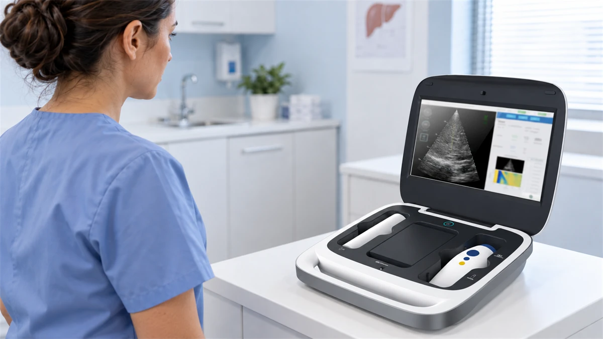
A liver elastography device provides non-invasive measurements related to liver stiffness. Some ultrasound systems also measure attenuation to support assessment of liver fat. The main technology groups are transient elastography, point and two-dimensional shear-wave elastography, and magnetic resonance elastography. Each combines a measurement method with a particular imaging and operating setup.
Choosing between them means understanding what the instrument measures, how the operator acquires a usable result, and how that result reaches the clinician. When comparing systems, examine the measurement method, validated thresholds, and cross-platform agreement alongside the units.
We developed Liverscan® around transient elastography with real-time B-mode guidance in a wireless probe. Its design and deployment examples illustrate how these technical distinctions affect everyday use.
What Is a Liver Elastography Device?
Elastography measures tissue response to a mechanical disturbance. In liver applications, the system tracks shear waves and estimates stiffness from their propagation. The resulting measurement contributes to the assessment of fibrosis, or liver scarring.
Stiffness is a physical property, and inflammation, congestion, and other conditions can also raise it. Clinicians therefore interpret elastography alongside medical history, laboratory findings, and other imaging when assessing fibrosis stage.
The EASL guidance on non-invasive liver tests explains these influences. For equipment selection, the consequence is straightforward: choose a system whose validated use and reporting fit the clinical decision it will support.
Main Types of Liver Elastography Technology
Transient elastography (TE) applies a controlled mechanical pulse through a dedicated probe. Ultrasound tracks the resulting shear wave. A dedicated TE system provides a focused liver-assessment workflow; the available anatomical guidance and attenuation measurements vary with the platform.
Point shear-wave elastography (pSWE) uses acoustic radiation force impulse (ARFI) technology. A focused ultrasound pulse pushes tissue, and the system measures shear-wave speed within a selected region. Integration with an ultrasound platform allows anatomical imaging during positioning.
Two-dimensional shear-wave elastography (2D-SWE) also uses ARFI excitation. Multiple pushes and rapid tracking produce a color-coded map of shear-wave speed or stiffness. The operator evaluates the map and quality information before placing a measurement region.
Magnetic resonance elastography (MRE) combines an external vibration source with magnetic resonance imaging (MRI) to map stiffness across sampled liver regions. It requires an MRI scanner, compatible equipment, and an MRI examination workflow. Consider it within an imaging or referral pathway rather than as a room-to-room clinic device.
For a department that already uses diagnostic ultrasound, compare the elastography package with current scanner compatibility and appointment capacity. For a dedicated liver service, price a stand-alone workflow. Compare quotations only after confirming the complete probe-and-software configuration, including the terminal, licenses, and accessories required for the examination.
What Does a Liver Elastography Device Measure?
Liver stiffness measurement (LSM) is the core output. TE platforms commonly report stiffness in kilopascals (kPa); pSWE and 2D-SWE systems may display meters per second (m/s), kPa, or both. Measurement ranges vary by system. Compare stated ranges together with validation data for the intended population and endpoint.
Ultrasound attenuation describes the loss of sound energy as it travels through tissue. Attenuation-based parameters can support steatosis assessment, meaning assessment of fat accumulation in the liver. Their names, algorithms, sampling methods, and validated interpretation differ across platforms. Interpret attenuation on the platform’s validated scale and reporting framework; direct liver-fat percentage is a separate measurement concept.
Obtaining stiffness and attenuation in one examination can consolidate part of the assessment into a single visit. A usable workflow keeps the two outputs distinct in the report and routes them to the appropriate review or referral step.
Before purchase, use an anonymized report to verify the measured quantities, units, quality statistics, and basis for any displayed categories. For longitudinal follow-up, a shared kPa unit is not sufficient evidence of interchangeability. Cross-platform changes are interpretable only when the device and acquisition method are documented and suitable agreement evidence is available.
How Imaging Guidance and Measurement Placement Differ
Anatomical imaging shows where the measurement will be taken. B-mode ultrasound provides a grayscale view of liver tissue and visible structures. A stiffness map displays the spatial distribution of elastography measurements. Signal indicators describe acquisition quality. These functions answer different questions during an examination.
B-mode-guided TE uses TE for excitation and tracking, with the anatomical image supporting sampling-location selection. In 2D-SWE, a different excitation and mapping method produces a spatial stiffness map.
In a hands-on trial, have the operator identify the target and adjust the region of interest (ROI). Ask what happens when a vessel, rib shadow, or poor signal affects placement. Observe how the operator recognizes an unsuitable acquisition and repositions the probe. Training and system-specific repeatability evidence remain important when assigning examinations to new staff.
Quality Control and Result Reliability
Review the complete measurement set rather than a single displayed value. The median summarizes the accepted readings. The interquartile range (IQR) describes the spread of the middle half; the IQR-to-median ratio expresses that spread relative to the median.
Historical TE protocols combined measurement count, variability, and success rate. Apply these criteria according to the relevant clinical guidance and device-specific instructions. Stiffness and attenuation may also use different quality summaries.
The Society of Radiologists in Ultrasound consensus addresses acquisition quality and reporting. In a hands-on evaluation, review failed attempts as well as successful examinations. During the trial, ask the operator to explain a rejection message, repeat the acquisition, and identify an inadequate data set without supplier assistance. Include a report with variable readings in the review. Check whether the summary preserves enough information for the interpreting clinician to understand the acquisition, including the measurement method, accepted count, and variability. If the record does not support a reliability decision, route the examination for repeat acquisition or clinical review.
How Device Format Changes the Clinical Workflow
Follow the examination through four stages: patient preparation, acquisition, quality review, and report handoff. A compact probe affects only part of that sequence.
|
Workflow stage |
What the service needs to establish |
|
Patient preparation |
A suitable examination space, preparation instructions covering fasting, positioning, and breathing, and a trained operator. |
|
Acquisition |
Working probe and terminal, positioning controls, and access to the required measurement mode. |
|
Quality review |
Visible acceptance criteria, a repeat procedure, and responsibility for unresolved results. |
|
Report handoff |
Correct patient identification, an agreed delivery method, and a clinician responsible for interpretation. |
Specialist liver clinics: Trace one follow-up patient through record retrieval, acquisition, quality review, and report filing. If broader abdominal imaging remains on a separate ultrasound platform, record the additional handoff and any duplicate data entry.
Primary-care and metabolic clinics: Before installation, assign a named owner for examination, quality review, referral, and report delivery. Include training and supervised practice in the launch plan. During the demonstration, run one appointment that requires repeat acquisition and report handoff. Accept the workflow only when staff can complete each step and identify the next clinical action without supplier prompting.
Community and mobile services need a workable transport, charging, cleaning, and report-delivery process. Test the complete setup at the intended location, including the terminal and any network-dependent handoff.
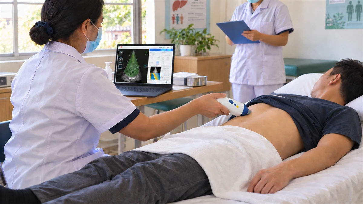
Research sites: Record the device, probe, software, preparation, and acquisition protocol for every research workflow. Review equipment or software changes before they enter ongoing data collection.
A community deployment shows why these responsibilities extend beyond the examination room. In Wencheng County, we donated Liverscan equipment to support a permanent township screening service launched in April 2024. Hospital specialists provided clinical support, and local authorities coordinated community participation. By July 2025, the program covered 13 villages. The deployment paired equipment with specialist support and local coordination.
How to Evaluate Clinical Evidence and Regulatory Fit
A biopsy-referenced study evaluates performance against tissue findings. A paired-device study examines how results relate across systems. Repeatability studies address consistency within or between operators. Keep those purposes separate when reviewing a supplier’s evidence.
Match the study population, clinical endpoint, and tested configuration to the proposed service. Read sensitivity, specificity, or discrimination results in relation to the question studied. Review performance separately for cirrhosis, significant fibrosis, and any other stage relevant to the proposed service.
Verify the local authorization, intended use, quoted model, and software configuration as one procurement workstream. Assess comparative clinical performance through studies that match the relevant population and endpoint.
How Liverscan Brings These Functions Together
Anatomical Targeting and Two Measurement Outputs
At the center of our system is a wireless composite probe that combines TE with real-time B-mode imaging. The operator can view anatomy and adjust the sampling position before acquisition. The design incorporates patented imaging-guided elastography technology. Depth and radial position can be adjusted through the probe controls, letting the operator move the sampling region away from visible large vessels or cysts.
During acquisition, our platform captures LSM and MAP, an ultrasound attenuation parameter reported in decibels per meter (dB/m). Both outputs belong to the same examination, while their clinical interpretation remains distinct.
In a sample Liverscan report, stiffness is summarized with a median and IQR-to-median ratio, while MAP is summarized with a median and IQR. This lets the clinician review each output with its corresponding variability information.
The clinical evidence for our B-mode-guided TE platform includes a 60-patient operator-reliability analysis published in 2024. Intraclass correlation coefficients, which describe measurement consistency, ranged from 0.824 to 0.913. Use this finding as repeatability evidence for the studied platform and operator conditions; assess the contribution of imaging guidance through the broader evidence set.
Acquisition Support and the Four-Measurement Research
Four artificial intelligence (AI) functions support our acquisition workflow: elastogram quality assessment, shear-wave trajectory detection, measurement-mode selection, and liver-boundary segmentation. These functions address defined acquisition tasks. During the demonstration, record accepted and rejected acquisitions, repeat attempts, operator interventions, and report completion with the quoted software; request clarification or a retest if the workflow cannot be reproduced.
A 2026 prospective study evaluated an AI-assisted point-of-care TE system developed on the Liverscan technology platform. It included a 138-patient biopsy cohort and a separate 1,455-patient paired-TE cohort. Research examinations collected ten valid measurements from the studied AI-assisted system.
An exploratory analysis compared medians derived from reduced measurement sets with the ten-measurement median. Four-measurement results were statistically non-inferior for the evaluated endpoints. The analysis used subsets from the same research acquisitions, with the ten-measurement median as the within-system reference.
In the biopsy cohort, stiffness achieved an area under the receiver operating characteristic curve (AUROC) of 0.97 for cirrhosis and 0.79 for significant fibrosis, stage F2 or higher. AUROC describes discrimination across thresholds. Because the single-center study used a prototype, confirm how the proposed commercial model and software relate to the studied system before adopting its reduced-acquisition protocol.
Two Configurations for Different Working Setups
We offer Liverscan C with a portable workstation and pre-installed software. Liverscan Mobile pairs the wireless probe with software for a compatible laptop or Surface. Both use the same probe platform. The workstation choice therefore belongs in the deployment assessment, alongside space, transport, and terminal management.
Results and reports are generated locally in our configurations, without cloud-based post-processing. The probe has a stated battery duration of 4+ hours. Assess probe battery, terminal endurance, charging, and report transfer together to establish the complete system’s operating time.
Match Liverscan C to service briefs that require a dedicated workstation. For Liverscan Mobile, confirm the institution’s computer and IT requirements before adding the configuration to the shortlist. In a model-specific evaluation, follow an examination from startup to report delivery with the proposed terminal. Include a room change or site transfer when the device will be shared.
For US procurement, our Liverscan Mobile is FDA-cleared under K233401, and Liverscan C under K242496. These records identify the individual models and their stated intended use. Check the authorization applicable to the destination market against the configuration being purchased.
Questions to Resolve Before Choosing a Liver Elastography Device
Use the proposed examination workflow to connect the technical and purchasing questions.
|
Area |
Question to resolve |
|
Technology |
Which method generates and tracks the shear wave? |
|
Outputs |
Which measurements, units, and quality summaries are included? |
|
Guidance |
What can the operator see and adjust during placement? |
|
Quality |
How are valid, rejected, and repeated acquisitions handled? |
|
Evidence |
Which patients, endpoints, and configurations were studied? |
|
Portability and deployment |
What must be transported, charged, cleaned, connected, and installed? Which terminal, software, power, and reporting arrangements are required? |
|
Regulatory fit |
Do the authorization, intended use, model, and software version match the destination market? |
Bring clinical, operator, and procurement representatives to the same demonstration. Mark each unresolved item as clarification required or retest needed, assign an owner, and record it against the quotation. Close the review only after the team can obtain a valid result, identify its quality status, and deliver the report to the intended recipient.
Request a model-specific Liverscan demonstration with our team. Share the care setting, operator experience, patient population, and reporting requirements so we can discuss a configuration suited to the planned workflow.
FAQs
1. How does a clinical program choose among transient elastography, pSWE, 2D-SWE, and MRE?
Match the modality to the clinical task and service setting. TE supports a focused point-of-care workflow; pSWE and 2D-SWE commonly operate within diagnostic ultrasound; MRE requires MRI infrastructure. Compare the exact configuration, imaging guidance, outputs, quality controls, evidence, reporting, and regulatory authorization. Clinical modality review
2. How accurate are liver elastography devices, and what factors affect measurement reliability?
No single accuracy percentage applies to every device or clinical use. Review the accepted measurement count, median, variability, rejection messages, and device-specific quality indicators. Match the supporting study to the intended population and endpoint; record any mismatch as an evidence gap. Interpret stiffness with relevant clinical information. RSNA consensus statement
3. Can liver stiffness results from different elastography devices be compared directly?
Not automatically. Shared units do not make results interchangeable because acquisition methods, algorithms, quality rules, and validated thresholds may differ. For follow-up, use the same validated system and protocol when practical. If the platform changes, document the model, software, and acquisition method, then review cross-platform agreement evidence before interpreting a change. Multi-device comparison study