
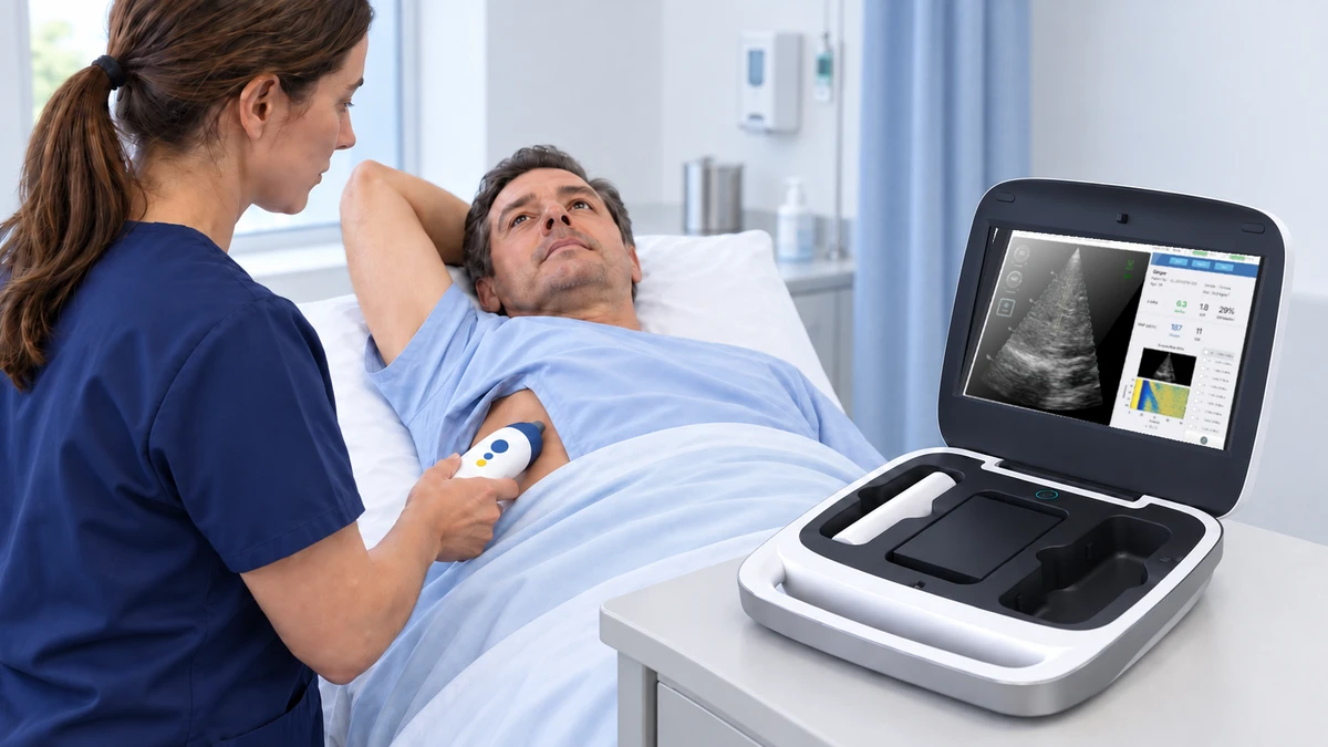
Using liver elastography for clinical trials requires more than a valid reading at a single visit. Changes in body position or preparation can complicate comparisons with baseline, even when individual examinations meet their quality criteria. Measurement quality and visit-to-visit comparability therefore need separate attention. Our Liverscan system combines real-time B-mode guidance with liver stiffness and ultrasound attenuation measurements in one examination. Guided targeting helps operators select a suitable region, while consistent procedures and documented acquisition conditions support interpretation across visits. Screening and follow-up measurements serve different purposes, shaping when results are collected and how they are interpreted. Across sites, separate quality checks for stiffness and attenuation and agreed rules for repeat scans help teams handle incomplete examinations consistently. Reports retain the acquisition conditions, deviations, and evidence limits needed to interpret an observed change.
Define the Measurement Role and Endpoint
Start by writing the question each measurement will answer. A screening value used to select participants has a different role from a follow-up value used to assess change. The distinction affects visit scheduling, repeat examinations, and the analysis.
Use the following framework to connect the measurement to a specific study decision.
|
Study purpose |
Decision to define |
Information to preserve |
|---|---|---|
|
Screening |
Which findings trigger further eligibility assessment? |
Screening value, method, and reason for referral |
|
Baseline stratification |
How are participants assigned to the planned groups? |
Accepted baseline examination and grouping rule |
|
Follow-up monitoring |
Is change expressed as an absolute difference, percentage, or trajectory? |
Baseline, visit timing, and results at each timepoint |
|
Treatment-response assessment |
What evidence supports the selected responder definition? |
Method, target population, and prespecified response rule |
Liverscan provides liver stiffness measurement (LSM), expressed in kilopascals, and MAP, an ultrasound attenuation parameter expressed in decibels per meter. As described in the 2026 Liver International study, MAP assesses ultrasound attenuation related to liver fat; it is not a direct percentage of fat.
We keep these outputs separate in the study plan. Assign each its own analysis role, quality assessment, and handling of missing results. When both are collected, a single appointment can serve both measurements without turning them into a combined endpoint.
Specify which examination establishes baseline and how an unscheduled repeat will be handled. Otherwise, two sites could choose different readings from the same visit and introduce inconsistency before follow-up begins.
Tie the measurement schedule to the comparison being made. When elastography is assessed against biopsy or another imaging method, define the permitted interval between assessments. For percentage-change analyses, identify the baseline value used in the calculation and retain the underlying measurements. A result collected outside the intended window needs an explicit analysis decision.
Make Repeat Measurements Comparable Across Visits
Keep the examination conditions recognizable
Several acquisitions within one examination describe the stability of that session. Measurements collected months apart also reflect changes in preparation, positioning, operators, and equipment. Same-session consistency and between-visit reproducibility therefore need separate attention, as demonstrated by the 2024 Radiology reproducibility study.
Build the visit around the same preparation instructions, body position, right-arm placement, breathing instructions, and anatomical window. Record the fasting interval and any departure from the planned conditions. The acquisition instructions should remain tied to the device instructions and study procedure, consistent with the QIBA Ultrasound SWS Profile.
When a participant arrives outside the preparation requirements, follow the agreed route: wait until the requirement is met, reschedule within the visit window, or document an allowed deviation. Collecting the scan without explaining the change leaves the later analyst unable to distinguish an unusual result from an unusual visit.
Use visible anatomy to support repeat targeting
As demonstrated in a 2024 Diagnostics comparison study, the real-time B-mode guidance in our Liverscan system shows the liver during targeting. Operators can identify a suitable region and avoid large vessels or non-liver structures.
At baseline, describe the measurement window and retain a reference image through the study’s image-handling process. At follow-up, use the recorded landmarks together with live anatomy. A previous image helps orientation; it does not guarantee that the same tissue has been sampled. This extends the guided liver elastography procedure into a longitudinal workflow.
Treat posture as part of the measurement
A 2024 prospective study using Liverscan assessed 31 healthy controls and 31 adults with chronic liver disease in supine, seated, and standing positions. Compared with supine measurements, average liver stiffness was higher in healthy controls and lower in participants with chronic liver disease when they sat or stood. Measurement success rates and the interquartile-range-to-median ratio, which describes the spread of readings, did not differ significantly across postures.
For repeat visits, we recommend keeping body position consistent and recording it alongside the measurement. When a participant is measured supine at baseline and seated at follow-up, document the change and apply the study’s predefined comparability rule. Passing the examination’s quality checks alone does not resolve the difference in acquisition conditions.
Separate technical change from clinical response
A technical-change threshold estimates whether a difference exceeds measurement variability under specified conditions. Use evidence matched to the system, measurement scale, and operator arrangement. A percentage derived from shear-wave speed cannot be copied directly to stiffness reported in kilopascals. (Radiology, 2024)
Prognostic associations ask whether measurements relate to future clinical events. A trial responder definition asks whether a participant meets a prespecified treatment-response criterion. These answer different questions. The 2023 Gastroenterology study on liver-stiffness dynamics provides prognostic evidence, but such an association cannot substitute for a technical-change threshold or a prespecified responder rule. Establish and validate each separately in the target population.
Site Consistency for Liver Elastography in Clinical Trials
A shared instruction sheet is useful only when sites can carry it out in the same way. Run a practice examination through the complete process: participant preparation, target selection, acquisition, recording, and transfer. Include an unsuccessful examination so staff can demonstrate what happens when an acceptable result is unavailable.
Manage access to previous results through the study’s blinding plan. Reference images can support positioning, while prior numerical values may need to remain hidden from the acquiring operator. Define that arrangement explicitly so follow-up staff at every site work with the same information.
Name the permitted device, probe, software version, and measurement modes in the site setup record. Training can then address the actual configuration used in the study. The Quantitative Imaging Biomarkers Alliance (QIBA) ultrasound measurement framework similarly assigns responsibilities across equipment, operators, quality assurance, and analysis. (QIBA Ultrasound SWS Profile)
Choose the configuration around the working environment
Both of our configurations use the same probe parameters. Liverscan C combines the wireless probe with a portable workstation; Liverscan Mobile pairs the probe with software installed on a compatible terminal.
The following deployment choices are practical planning recommendations.
|
Site arrangement |
Configuration to consider |
Setup decision |
|---|---|---|
|
Dedicated research room or shared examination station |
Liverscan C |
Allocate the workstation and define responsibility for keeping its configuration consistent. |
|
Bedside visits or sites using compatible laptops or Surface devices |
Liverscan Mobile |
Select a terminal meeting the software requirements and manage its installation throughout the study. |
A portable setup still needs an appropriate examination position, trained staff, and a defined route for transferring results. Avoid changing a participant’s measurement conditions simply because the probe can move between rooms.
Keep equipment changes visible
When an operator, terminal, probe, or software version changes, document the date and affected examinations. Repeat the relevant training or equipment checks, then assess whether the change could affect comparability. A switch to another elastography platform needs its own assessment; reporting the same unit does not establish interchangeable measurements. (Radiology, 2024)
Assign one contact at each site to resolve configuration questions. This keeps an unexpected replacement or update from becoming an undocumented change across several visits.
Trace a practice examination from the acquisition terminal to the study record. Check that participant codes, units, parameter labels, and visit identifiers survive the transfer. Include a corrected record in the exercise, with the original retained and the correction explained. This tests the handoff that a successful scan alone cannot demonstrate.
Turn Quality Checks Into Clear Decisions

Quality control works best when operators know the next action. Separate an invalid acquisition, an incomplete examination, and a visit performed outside the study conditions. Each calls for a different response.
Assess stiffness and attenuation independently
During acquisition, our four artificial intelligence (AI) models support elastogram quality assessment, shear-wave trajectory detection, measurement-mode selection, and liver-boundary segmentation. Acquisitions that the embedded algorithms classify as invalid are flagged and excluded by the system; study-level acceptance still follows the prespecified quality-control plan.
In the published study of AI-assisted point-of-care transient elastography (AI-POC-TE), MAP quality checks used liver-tissue segmentation independently of LSM’s elastogram assessment. (Huang et al., 2026)
Carry that distinction into the study record. Give LSM and MAP separate quality status fields. When only one parameter meets its acceptance criteria, retain its status and process the other as unsuccessful. Whether the usable parameter enters analysis follows the prespecified plan; neither an automatic whole-exam rejection nor an automatic pass for both is appropriate.
Connect each problem to an action and a record
Use the following decision table as a starting point for site-specific instructions. Fill in the acquisition count, attempt limit, and visit window from the applicable device instructions and study plan.
|
Trigger |
Immediate action |
Record to retain |
|---|---|---|
|
Preparation or posture differs from the planned visit |
Correct the condition where feasible; otherwise use the rescheduling or deviation route. |
Actual conditions, reason, and disposition |
|
Targeting is unsuitable or an acquisition is flagged invalid |
Reassess the window, reposition as needed, and repeat within the attempt limit. |
Attempts, available quality flags, and outcome |
|
The examination reaches the stopping limit without acceptable results |
End the session and follow the rescan or escalation pathway. |
Failure reason and planned next step |
|
LSM and MAP have different quality outcomes |
Handle each parameter separately under its analysis rule. |
Result and quality status for each parameter |
|
Equipment changed or comparability remains unresolved |
Refer the examination to the designated study lead for a documented decision. |
Configuration, affected visit, and decision |
An unexpectedly high or low result alone is not a reason to keep scanning. Apply the same repeat criteria across treatment groups and visits. Retain the reason for each repeat and identify which examination supplies the analysis value. This keeps a reassuring-looking replacement result from silently displacing an earlier valid examination.
Put the four-measurement finding in context
In an exploratory analysis of the 2026 Liver International study, the AI-POC-TE prototype developed within the Liverscan framework produced four-measurement results reported as statistically non-inferior to ten for the evaluated diagnostic tasks. (Huang et al., 2026)
That finding informs acquisition planning. It does not establish a universal stopping rule for serial trial measurements. Set the count for the selected system and study purpose, then apply it consistently across sites and visits.
Track unsuccessful examinations by site, operator, and parameter. Repeated targeting failures call for a different investigation from missed preparation requirements or missing attenuation results. Look for the recurring cause, address the relevant training or setup issue, and record the action. Keep these quality checks separate from treatment-response results so that apparent improvement never becomes an acceptance criterion.
Report Results and State the Evidence Limits
A useful report lets another study team member understand how the result was obtained and why it entered the analysis. Keep the participant identifier, site, operator, visit date, device configuration, acquisition conditions, parameter-specific results, and quality status together.
Add the reason for missing data, repeats, and deviations. These are study records: some may come from the instrument, while others require site documentation. Preserve the original result when a later assessment changes its analysis status.
Distinguish a missed appointment from an attempted examination that failed, and distinguish both from a valid measurement excluded because its conditions differed from the study plan. Use separate status fields instead of one blank value. The analysis team can then apply the planned missing-data and sensitivity analyses without guessing what happened at the site.
The clinical evidence behind our platform also needs precise description. The 2026 study included a 138-patient biopsy cohort and a separate 1,455-patient paired-elastography cohort. The former evaluated diagnostic performance; the latter demonstrated correlation with the comparator system. These findings establish neither interchangeability nor longitudinal endpoint validity. (Huang et al., 2026)
The clinical evidence for Liverscan connects these findings with the published investigations. Match the population, system, and research question to the intended use rather than treating the total participant count as validation of every application.
For an unexpected follow-up change, present the measurement alongside the visit conditions and relevant clinical findings. Lower stiffness alone does not demonstrate fibrosis regression because factors beyond fibrosis can affect stiffness (BMC Gastroenterology, 2024). Report the observed change first, then explain the interpretation supported by the study design. Where comparability remains uncertain, retain that uncertainty in the result.
Frequently Asked Questions
Can participants be measured while seated?
The posture study demonstrated seated measurement with Liverscan (BMC Gastroenterology, 2024). A study using that position needs a consistent positioning plan and an appropriate interpretation framework. Avoid pooling seated and supine follow-up measurements as though posture were unchanged.
How long does a research visit take?
Allow separately for preparation, positioning, acquisition, any permitted repeats, and documentation. Time the complete workflow during site setup. Acquisition speed alone is a poor basis for booking visits or estimating staff workload.
What happens when an examination produces no usable result?
Record the attempted visit and the specific failure reason. Apply the planned rescan window or missing-data rule. A blank result with an explanation is more informative than an unrecorded failed examination.
Summary
Reliable serial liver elastography depends on more than obtaining a valid value. The endpoint role, visit conditions, site configuration, parameter-level quality control, and reporting rules must remain aligned from baseline through follow-up.
To discuss a Liverscan clinical-trial workflow, share the target population, measurement roles, visit schedule, and planned site setup with our team. Include any defined rules for repeat measurements and rescans, along with the required data outputs.